We never know how someone has their bike set up? There is much behind what we do! We don't just eye things!
Why in other sports like golf do people go and spend so much time at the driving range to practice their swings. Why do they go to the practice green to work on their putting. Why do they go to a PGA pro to learn? Tons of time working on the small things like their grip, addressing the ball, getting the club to make perfect contact. They work very hard on their take-away and their timing to obtain better results.
Money can buy the very best clubs (bikes) you want, but you still have to learn how to use them. Infact you can spend a life time and never learn the swing! Sure you enjoy the game, posting high scores. Ha!
Then you take the cyclist who spends much money on their $$$$ bike, this and that, a coach for training, mostly online. In most cases, its a "training coach" doesn't live near you to watch or know how you pedal? How would they, if they don't have sEMG and Dartfish working together? Just using Dartfish is not letting you know the tone of the muscles.
That is the whole point of Myo-facts sEMG/Dartfish, That is why we have taken over two years now, putting them together!!!
We don't think a bad golf stroke is going to imporve your game. The same holds true about a bad pedal stroke, how is it going to improve your game? A powermeter helps in training, but only records the sum of the muscles and it will not show you the loads on your muscle! It will not inform you or the coach on what muscle is doing what!
It seems most do their thinking from "hear-say', or they read something in a mag, or not at all? They just jump on their bike and ride "Bad motor skills" or not.
Myo-facts sEMG/Dartfish is a major break through and it really makes a difference for learning. It shows you the "TRUTH". Read whatever, study whatever, but the bottom line is your "Know How". Where you get your "Know How" makes a difference!
But first understand this, if you can't address the club head on the ball, good luck. Every stroke counts, and every pedal stroke counts.
Top coaches like (Hunter Allen, Rick Crawford, Kendra Wenzel, etc...) are sending their racers our way! They all know that we provide the best fit and biofeedback, leading to better results.
Why should you care about your pedal stroke? The person who wants to live a long active life style!

The knee joint as a mechanism of three joints (Biomechanics).
The knee joint is a complex mechanism of four bones the femur, tibia, patella and fibula which interact in three separate joints the tibiofemoral, patellofemoral, and tibiofibular joint. The function of these joints is to allow certain movements, restrict others, and to provide load transfer through the lower limb.
Tibiofemoral joint loading: tensile and compressive forces (Biomechanics).
The knee is subjected to high external forces in many directions: compression, anterior-posterior shear, medial-lateral shear, internal-external, flexion, and varus-valgus moments. The ligaments and other soft tissues, such as the posterior capsule (image) , hold the knee joint together. These tissues are under great tension with each pedal stroke. The articular surfaces and the menisci hold the knee joint apart. These are under compression with the pedal stroke. Joint loading is estimated by applying the results of pedal analysis to a joint model. The tibiofibular joint is loaded to approximately three times body weight during walking, with most of the compressive force passing into the medial compartment. It is less when on the saddle, but still subjected to high external forces.
The knee joint is the largest synovial articulation in the body, and of all joints possesses the most voluminous synovial cavity. It is defined as a compound joint on account of the irregularity of contour, and the number, of its articular surfaces. Correspondingly, movements of the knee joint are complex in the pedal. Thus the knee joint is capable not only of flexion and extension, but also of "complex rotatory movements".
The knee joint is also described as a complex joint because of the presence of intraarticular menisci and intraarticular ligaments. Even with the perfect fit, the tone and timing of the quads changes its loads and functions.
The knee joint is an articulation between the lower end of femur, the meniscus-bearing upper surface of tibia, and posterior surface of the patella. Although sharing a single capsular investment and a single synovial cavity, the knee joint is a complex of three articulations; a medial tibio-femoral articulation, a lateral tibio-femoral articulation and a patello-femoral articulation.
In terms of function and movement the knee joint may be defined as a modified hinge joint, capable of flexion, extension and rotation.
Despite the apparent lack of congruence between the various articulating surfaces, the knee joint is a remarkably stable joint. This stability is due largely to the presence of several strong ligaments (both intracapsular and extracapsular), and various tendinous and muscular attachments.

Medial tibial plateau (Biomechanics)
The concave medial tibial plateau increases knee joint stability when under compression, as the resultant compressive forces act to locate the medial femoral condyle in the concavity.

Lateral tibial plateau (Biomechanics).
The convex or flat lateral tibial plateau allows more mobility of the lateral compartment of the knee. Therefore, the motion of the lateral compartment is guided primarily by the muscle loading, soft tissue restraints, and external moments, rather than the joint contact force between the femoral condyle and the lateral tibial plateau. The only way to see the muscle loading is by using EMG.
This mobility of the lateral compartment is more pronounced with injury to the soft tissue structures of the knee which restrict tibial rotation, such as the anterior cruciate ligament. Such an injury frequently causes the 'pivot shift', which is the movement of the tibia from one position of stability to another. Bad motor skills also decrease its stability.

Even with the presence of the several strong ligaments and tendinous and muscular attachments poor loading of certain muscles with a poor pedal stroke/saddle location can cause mobility issues at the knee.

Patellofemoral joint (Biomechanics).
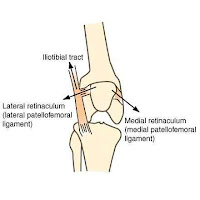
The patellofemoral joint has six dof. The patella is very free to move passively, that is, the ligamentous restraints to patellofemoral motions are small when still, but under active motion the patella follows a reproducible path of motion. The motion is known as the patellar tracking and a measure of the freedom-of-motion is known as the patellar laxity, or patellar stability.
The patellofemoral joint has one main mechanical function: to provide a fulcrum to increase the lever arm of the quadriceps to extend the knee joint, or to stop it from flexing. These are critical in all aspects of cycling, as is only takes milimeters to be off. The extensor force required for this activitie is very high. The force magnitudes across the whole extensor mechanism of the knee are thought to be the highest in the whole human body. The hyaline cartilage of the patella is the thickest in the body and provides an environment with minimal frictional losses under high compressive loads.

Physiological cross sectional area (Biomechanics).
Muscle strength and loading is difficult to quantify as direct measurement of muscle forcesl. A number of different approaches for quantification of muscle forces have been applied in the literature. Electromyography can indicate the intensity of muscle activation, but it is not yet possible to determine accurately the force exerted by each muscle in a complex. One approach is to relate a muscle's ability to generate force to its size and architecture.
Cross sectional area has been used to measure force ratios. The physiological cross sectional area (image) (PCSA), (cross sectional area of the muscle perpendicular to all of its fibers), is however believed to provide a estimation of muscle strength, being proportional to the number and cross sectional area of the tension-producing fibers.
The PCSA can indicate the contribution of each muscle in a group of muscles, particularly when they reach a limiting stress, such as in strenuous activity like cycling. This is where we take our readings!


Patella (Biomechanics).
The patella is the largest sesamoid bone in the body and has the thickest hyaline cartilage in the body. The areas of the medial and lateral facets reflect the ratio of patellar joint force that they transmit: the Q-angle biases the load 60:40 onto the lateral facet.
The most obvious morphologic feature of the articular surface of the patella is its asymmetry. This is important as very few people have the same shape. This morphologic differences produces different pedal styles. We do not want to change one's style to match another for this reason. There are many simply using lasers to keep everything on the same plane. This is wrong!
Based on this asymmetry (Wiberg) classified patellae into three types:
Type I: the medial and lateral facets are gently concave and nearly equal in size.
Type II: the medial facet is flat or convex and smaller than the concave lateral facet.
Type III: the medial facet is very small and is usually prominent and convex, while the lateral facet is broad and concave.
The most common type is type II (55%), while type III accounts for 25%. Other types of classification systems have been proposed, the most common is the one proposed by Ficat and Hungerford (1977) which is based on the angle subtended by the two major facets.
The primary function of the patella is to increase the lever arm of the extensor muscles. This requires a surface adapted to bearing high compressive loads with minimal friction. This can change, given one's focus on the firing of their extensor muscles.

In our fitting process, we don't just use a plum-line to find a saddle location. Even the best of pros are learning from our EMG, as they have a better chance of firing their muscles at the best time. More important, they stay in the game!
Their is much more to our fit than meets the eye.
After we find your constraints, we use a very powerful CAD program in the background of our site to get it right! Then we use the Myo-facts sEMG/Dartish with great care to find the combination of medial/lateral & flexion/extension loads for the type of game you wish to play.
What's in your game?


No comments:
Post a Comment